When photographers take long-exposure photos, they maximize the amount of light their camera sensors receive. The technique helps capture scenes like the night sky, but it introduces blurring in the final image, as in the example at right.
It’s not too different from cryo-electron microscopy, or cryo-EM, which scientists use to study the structure of tiny molecules frozen in vitreous ice. But while motion-induced blur in photography can create beautiful images, in structural biology it’s an unwanted side effect.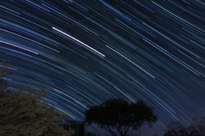
Protein samples for cryo-EM are frozen at -196 degrees Celcius to protect the biological structures, which would otherwise be destroyed by the microscope’s high-energy electron beam. But even when frozen, samples are disturbed by the powerful electron dose, causing motion that would blur a long-exposure photo.
To get around it, UCSF researchers use specialized cameras to instead capture videos of the biological molecules, so they appear nearly stationary in each frame of the video. Correcting the motion across frames is a computationally demanding task — but can be done in seconds on NVIDIA GPUs.
“If the motion was left uncorrected, we’d lose the high-resolution picture of a molecule’s 3D structures,” said Shawn Zheng, scientific software developer at the University of California, San Francisco and Howard Hughes Medical Institute. “And knowing the structure of a molecule is critical to understanding its function.”
Zheng and his colleagues run MotionCor2, the world’s most widely used motion-correction application, on NVIDIA GPUs to align each molecule in the video from frame to frame — creating a clean image researchers can turn into a 3D model.
These 3D models are essential for scientists to understand the complex chains of interactions taking place in an individual protein, such as spike proteins on the COVID-19 virus, speeding drug and vaccine discovery.
Solving the Bottleneck
UCSF, a leader in cryo-EM research, has been the source of groundbreaking work to improve the resolution of microscopy images. The technology enables scientists to visualize proteins at an atomic scale — something considered impossible just a decade ago.
But the pipeline is lengthy, involving freezing samples, capturing them on multimillion dollar cryo-EM microscopes, correcting their motion and then reconstructing detailed 3D models of the molecules. To keep things running smoothly, it’s critical that the motion-correction process runs fast enough to keep pace with the new data being collected.
“Cryo-EM microscopes are very expensive instruments. You don’t want it just sitting there idle. But if we have a backlog of movies piled up in the machine’s data storage, nobody else can collect more,” said Zheng. “It’d be a waste of this expensive instrument, and slow down the research of others.”
To achieve rapid motion correction, UCSF’s Center of Advanced Electron Microscopy uses workstations with eight NVIDIA GPUs for each microscope. These workstations are needed to keep up with the cryo-EM data collection, which acquires four movies per microscope per minute.
The GPU setup can run eight jobs concurrently, taking on the iterative process of motion correction for videos with as many as 400 frames, each with nearly 100 million pixels.
To speed the development of new applications, Zheng, who’s used NVIDIA GPUs for his research for a decade, uses a workstation powered by two NVIDIA Tensor Core GPUs. The system can analyze a 70GB microscope movie in under a minute.
Accelerating COVID Research
Zheng and his colleagues also use GPUs to run alignment software for cryo-electron tomography, or cryo-ET. This technique is better suited to study slightly heterogeneous specimens like macromolecules and cells. Samples are tilted at different angles, collecting a series of images that can be aligned and reconstructed into a detailed 3D model.
NVIDIA GPUs can fully automate the reconstruction process, taking a half hour on a single GPU, he says.
In a recent paper in Science, Zheng collaborated with lead researchers from the Netherlands’ Leiden University Medical Center to use cryo-ET to study molecular pores involved in COVID-19 virus replication in cells. A better understanding of this pore structure could help scientists develop a drug that targets it, blocking the virus from replicating in an infected patient.
To learn more about Zheng’s work, watch this on-demand talk from the GPU Technology Conference.
Main image shows a cryo-EM density map for the enzyme beta-galactosidase, showing the gradual increase in quality of the cryo-EM structures from low to high resolution. Image by Veronica Falconieri and Sriram Subramaniam, licensed from the National Cancer Institute under public domain.
The post Freeze the Day: How UCSF Researchers Clear Up Cryo-EM Images with GPUs appeared first on The Official NVIDIA Blog.